
Chinese researchers grew the sinoatrial node, which initiates the heart’s rhythm, in a laboratory environment. The three-dimensional organoid developed with human pluripotent stem cells can beat rhythmically on its own, generate electrical signals and interact with nerve cells, offering a new model for heart rhythm research.
Biological pacemaker that manages heart rhythm was moved to the laboratory
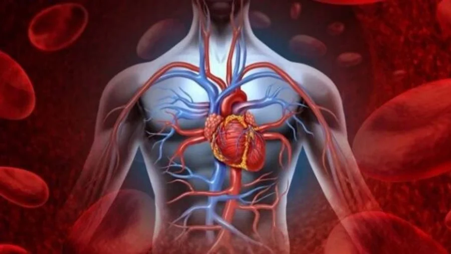
In the study conducted in Shanghai, researchers developed an organoid that mimics the sinoatrial node, which is located in the right atrium of the heart and serves as a natural pacemaker. The sinoatrial node initiates electrical impulses in the heart, and these impulses determine the contraction pattern of the atria and ventricles. When a problem occurs in this structure, the heartbeat can become dangerously slow, become irregular, or short-term pauses may occur.
The study was conducted by Prof. from the Center for Excellence in Molecular Cell Science, affiliated with the Chinese Academy of Sciences. The team led by Zeng An and Prof. from Fudan University. Luo Zhe and Prof. from Tongji University. Prepared with the contributions of Du Meirong.
Researchers created three-dimensional sinoatrial node organoids by manipulating human pluripotent stem cells with specific developmental signals. These cells gave rise to structures containing pacemaker cell types corresponding to the head, tail, and transition regions of the native sinoatrial node.
The developed organoids were able to beat rhythmically on their own. In the study, it was reported that these structures carry gene expressions similar to human embryonic pacemaker cells and respond to drugs that change heart rate. It was observed that electrical signals were transmitted outward from the sinoatrial node when the organoids were connected by atrium-like structures. Thus, the basic “stimulus production and transmission” process in the heart was modeled in a laboratory environment.
The team also tested whether the system could be used in disease modeling. When a mutation linked to familial slow heart rhythm was introduced into the organoids, the biological pacemaker began beating more slowly.
In ongoing experiments, it was reported that the rhythm partially normalized with a selective potassium channel blocker. This phase revealed that the model could be used to study the mechanism of heart rhythm disorders and evaluate possible treatment candidates.
One of the important parts of the research was the nervous system connection. Heart rate is regulated in the body not only by the sinoatrial node but also by the autonomic nervous system. Therefore, researchers developed cardiac plexus organoids containing parasympathetic-like neurons and combined them with pacemaker organoids. In the experiments, it was noted that the nerve fibers extended towards the pacemaker cells and reduced the organoid’s beating rate.
Later, atrium-like organoids were added to the system and the “nerve-sinoatrial node-atrium” structure was established. It was stated that in this triple assembloid structure, the neural regulation signal does not only remain in the pacemaker region, but is also transported to the downstream atrial tissue. The research provides new experimental ground for studying how rhythm in the human heart is fine-tuned by the nervous system.
The study also examined a molecular pathway related to the maturation of pacemaker cells. In spatial transcriptomic analysis, it was determined that the GPR37 receptor was found in pacemaker cells and the PSAP ligand was found in neighboring neurons.
In experiments, it was reported that PSAP supports the maturation of pacemaker cells through GPR37, and when this signal is disrupted, maturation is weakened, and when PSAP is added, maturation signs return.


Comments
You can write your views about this story. Comments may be moderated according to site settings.